


Imaging is vital in the diagnosis and management of thyroid cancer. The doctors may make many treatment decisions based on what they see on the diagnostic scans, and many types of scans are available. An overview of each is described below, and more detail discussions are available by following the appropriate link.
These diagnostic scans may include:
Radioiodine whole body scanning for differentiated thyroid cancer (e.g., papillary and follicular) is a valuable diagnostic imaging tool. The physician may use this type of scanning to help evaluate, manage, and monitor these types of thyroid cancers.
A radioiodine scan involves the oral administration of a small amount of radioactive iodine. The iodine is like the iodine that’s added to most table salts. The difference, however, is that the iodine is “radioactive,” which means that it emits a small amount of energy that can be imaged with special cameras. We emphasize that the amount of energy released in this process is small and, in most cases, negligible.
Several hours to one or two days after the administration of the radioactive iodine, images are obtained of the whole body using a special camera to look for any thyroid tissue that takes up this small amount of radioactive iodine. Thyroid cells take up iodine because iodine is needed for the thyroid cells to try to make thyroid hormone.
An image typically takes 15 to 45 minutes to obtain, depending on the location; often, multiple images are obtained. Longer imaging times and multiple images help maximize the information obtained from the scan and helps ensure that any treatment is individually tailored to each patient’s specific situation.
Although some facilities will not routinely perform radioiodine scanning prior to any radioiodine therapy, MedStar Health routinely performs radioiodine scanning prior to any radioiodine therapy. In addition, these scans can be performed in many different ways, and our staff will schedule each patient for the scanning technique that is most appropriate for their situation.
In preparation for a radioiodine scan, our team will consider a variety of factors, such as withdrawal of thyroid hormone vs Thyrogen® injections, low-iodine diet, and/or other special preparations. Patients will also receive our “Six-Step Instructional Binder,” which will help guide patients through the preparation steps.
Patients will also receive a complimentary copy of “Thyroid Cancer: A Guide for Patients,” published by Keystone Press, which may also be purchased from the Thyroid Cancer Survivors Group, Inc. (ThyCa) online at www.ThyCa.org and Amazon.com.
A positron emission tomography-computerized tomography (PET-CT) scan is an advanced, non-invasive test that uses an additional imaging tool to detect tumors. This imaging technique can sometimes detect tumors earlier and more reliably than other imaging tools.
A PET-CT scan involves the injection of a small amount of radioactive sugar called FDG (fluorodeoxyglucose) into a patient’s vein; images of the whole body are obtained. Because tumor cells are typically very active and need sugar, they take up the radioactive sugar. This allows the PET-CT camera to image the degree of radioactivity.
The CT scan is done by a second imaging camera that shows the anatomy. An analogy to use to understand these two imaging techniques is a car engine. The CT obtains images of the car engine’s anatomy, while the PET obtains images of the function of the car engine’s anatomy. The PET and CT pictures are taken essentially at the same time and the combination of both provides better diagnostic accuracy.
MedStar Health was the second of two institutions in the US to initiate the combined use of a PET and CT scan. This technique is an advance in quality that allows for faster and better images, as well as lower doses of the radioactivity. Better images provide better localization of the cancer sites, which helps physicians to better individualize each patient’s treatment plan.
As already noted, computed tomography (CT) or computed axial tomography (CAT) scans obtain excellent images of the patient’s anatomy. Images may be obtained at the same time as the PET images or obtained at a different time or day.
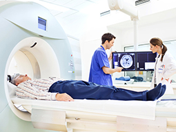
Magnetic resonance imaging (MRI) is a valuable imaging tool. MRIs use very strong magnets that align atoms in the various organs. When the magnet is turned off and the atoms go back to their original alignment, radio signals are emitted. With a special machine, the radio signals are gathered and images are obtained.
Just as PET and CT scanners have been combined into a single imaging unit, PET and MRI scanners have also been combined into a single imaging unit. Few of these combined scanners are available; however, with the use of computer software, the images obtained with the PET scanner may be electronically overlaid with the images obtained with the MRI scanner. The combining of two different imaging technologies (e.g., fusion) provides superior quality images that allow better localization of any cancer site.
Dosimetry is a combination of specialized pretherapy radioactive iodine scans and measurements that allow physicians to calculate the maximum dose of radioactive iodine that can be given to a patient for a therapy. This may be requested for local thyroid cancer remaining in the neck or for thyroid cancer that has spread to a location outside of the neck.
Our center is one of the few in the US to use this specialized measurement to help maximize the therapeutic effect to a patient’s thyroid cancer while helping to ensure that the patient does not receive an excessive amount of radioiodine, reducing the likelihood and severity of any side effects to the patient’s bone marrow from the radioactive iodine.
Because some patients travel long distances to our facility and/or their schedule does not allow them to participate in the standard dosimetry scans and measurements described above, we offer a modified and significantly shortened method, called a simplified dosimetry measurement (or percent 48-hour whole body retention).
Like dosimetry, this simplified measurement helps to determine the maximum dose while also helping to ensure that patients do not receive an excess amount of radiation exposure to their bone marrow.
Our providers
Expert radiology care
Getting the care you need starts with seeing one of radiologists.