


In the fight against disease, diagnosis is critical to the best treatment and outcome. At MedStar Health, we use nuclear medicine imaging techniques to give doctors and staff a unique way to look inside the human body—and make sure you are getting the best care.
Nuclear medicine tests use small amounts of radioactive material to painlessly diagnose or treat conditions in the body. These test require a material called a radiotracer. Depending on the type of study that you are having, this tracer is injected into a vein, swallowed, or inhaled. It gathers in the area of your body that is being examined and gives off energy in the form of gamma rays.
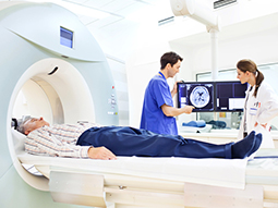
Most nuclear medicine procedures use a gamma camera, which finds the gamma rays (radiation) and take pictures from different angles.
Gated Perfusion Imaging
Gate perfusion imaging evaluates motion of the heart muscle and determines how well it is pumping. We perform this test at the same time as myocardial perfusion imaging (testing how well blood flows through your heart), using a special computer program.
By doing these tests at the same time, we are able to evaluate both blood flow and pumping function of the heart in one study and, in the majority of cases, in one visit. The results of gated perfusion imaging provide important information on prognosis and risk in patients with coronary artery disease.
Multiple Gated Acquisition (MUGA)
MUGA evaluates heart muscle motion and pumping function. The test is ordered primarily in patients undergoing chemotherapy to determine the presence and extent of side effects of chemotherapy on heart function. Patients with heart failure or heart valve disease also can be evaluated with this study to determine the severity of the disease.MUGA uses a radioactive tracer that attaches (or tags) to the red blood cells circulating through the heart. Scans of the heart are also synchronized with the patient's EKG for evaluation of heart muscle motion and pumping function of the heart.
Myocardial Perfusion Imaging (MPI)
MPI uses a radioactive tracer to evaluate blood flow to the heart muscle. While cardiac CT angiography and cardiac catheterization diagnose how serious coronary artery disease may be, MPI diagnoses the effect of the coronary artery disease on the blood flow to the heart muscle. Insufficient blood flow to the heart muscle causes chest pain (ischemia) experienced by patients with significant coronary artery disease.MPI is performed in two parts:
Rest Study: In the rest study, the patient is injected with a radioactive tracer in a vein. Depending on the type of radioactive tracer used for the rest study, the patient then waits approximately 30 to 45 minutes. Pictures of the heart are taken with a special type of camera, called SPECT.
Stress Study: During the stress study, the patient undergoes a stress test on a treadmill. If the patient cannot exercise on the treadmill, a drug is given through an IV that simulates stress. A cardiologist monitors the patient's blood pressure, heart rate, and EKG throughout the stress study. At a specified time during the stress test, the patient is injected with the radioactive tracer and, shortly thereafter, the stress test ends.Depending on the type of stress test performed and radioactive tracer injected during the stress study, the patient waits approximately 15 to 30 minutes before scans of the heart are taken with a SPECT camera.Two types of SPECT cameras are available:
- Conventional SPECT camera: With the conventional SPECT camera, the patient lies flat on an imaging table, and the camera rotates around the patient's chest to scan the heart for approximately 20 to 30 minutes.
- New D-SPECT® camera: With the new D-SPECT® camera, the patient sits in a specially designed reclining chair with a smaller-sized camera that is fixed in place over the patient's chest. Unlike the conventional SPECT camera, the D-SPECT® does not rotate around the patient. The D-SPECT® takes scans of the heart for two to four minutes.
Our providers
Expert radiology care
Getting the care you need starts with seeing one of our radiologists.